New Patients: (561) 609-6491
Existing Patients: (561) 395-0550
7280 W Palmetto Park Rd
Suite 206N
Boca Raton, FL 33433

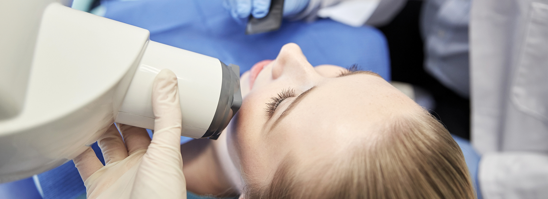
Digital radiography utilizes computer technology and digital sensors for the acquisition, viewing, storage, and sharing of radiographic images. It offers several advantages over the older traditional film based methods of taking x-rays. The most significant of these advantages is that digital radiography reduces a patient’s exposure to radiation. Other benefits are that images can be viewed instantly after being taken, can be seen simultaneously as needed by multiple practitioners, and can be easily shared with other offices. Digital x-rays are also safer for the environment as they do not require any chemicals or paper to develop.
An electronic pad, known as a sensor is used instead of film to acquire a digital image. After the image is taken, it goes directly into the patient’s file on the computer. Once it is stored on the computer, it can be easily viewed on a screen, shared, or printed out.
Among the most advanced digital x-ray technology available today, the Sirona Orthopos SL 3D Scanner offers the widest range of options and the best image quality to ensure accurate case diagnoses and to support complex treatment planning. This unit exposes patients to the lowest amount of radiation while obtaining the sharpest and most diagnostic images. The Sirona Orthopos SL 3D scanner provides the sophisticated images needed for implant procedures, airway analysis, endodontics, oral and maxillofacial surgery procedures, TMJ treatment, as well as general dentistry.